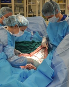
CALEC surgery, or cultivated autologous limbal epithelial cell surgery, represents a groundbreaking advancement in the field of eye damage treatment. This innovative procedure, performed at Mass Eye and Ear, utilizes stem cell therapy to restore the corneal surface, offering new hope to patients suffering from severe corneal injuries such as chemical burns and infections. By transplanting healthy limbal epithelial cells extracted from the patient’s unaffected eye, the surgical technique not only promotes healing but also enhances visual acuity, with a remarkable success rate of over 90% in clinical trials. Ula Jurkunas, a leading expert in ophthalmology, emphasizes the significance of CALEC surgery in transforming the quality of life for individuals facing previously untreatable conditions. With ongoing research and potential future applications, CALEC surgery stands at the forefront of corneal restoration therapies, paving the way for improved outcomes in eye care.
Cultivated autologous limbal epithelial cell transplantation, often referred to as CALEC surgery, is a revolutionary treatment aiming to repair corneal surface damage through the use of regenerative medicine techniques. This approach addresses limbal stem cell deficiency, a critical condition affecting patients with extensive eye injuries that traditional treatments struggle to resolve. By harnessing the power of stem cell therapy, specifically by harvesting healthy limbal epithelial cells from one eye, CALEC surgery facilitates the repair of the damaged eye, thus restoring its functionality and reducing pain. As this groundbreaking technique gains momentum, it showcases the incredible potential of modern ophthalmic innovations in addressing eye health challenges. With support from esteemed institutions like Mass Eye and Ear, CALEC could soon become a standard treatment option for patients in dire need of effective eye damage treatment.
Introduction to CALEC Surgery
The field of ophthalmology is entering a transformative era with the introduction of CALEC surgery, which offers new hope for patients with corneal damage. At Mass Eye and Ear, researchers have pioneered a revolutionary stem cell therapy that utilizes cultivated autologous limbal epithelial cells (CALEC) to restore the corneal surface in individuals suffering from limbal stem cell deficiency. This innovative surgery involves harvesting healthy stem cells from a patient’s unaffected eye and transplanting them to the damaged eye, aiming to rejuvenate the cornea that is often deemed irreparable.
With promising outcomes from clinical trials, CALEC surgery stands out as a beacon of hope for those battling blinding corneal injuries. Ula Jurkunas, the principal investigator, has highlighted that the treatment has shown over 90 percent effectiveness at restoring corneal surfaces. This milestone not only challenges the notion of untreatable corneal damage but also signifies a major advancement in regenerative eye care that could improve the quality of life for many patients.
The Process Behind CALEC Surgery
CALEC surgery involves a meticulous process that begins with a biopsy of the healthy eye to extract limbal epithelial cells. These cells are then cultured to create a cellular tissue graft, which typically takes two to three weeks. Once the graft is ready, it is carefully transplanted into the damaged eye, where it can integrate and potentially restore the cornea’s natural function. This complex procedure showcases the marriage of cutting-edge science with practical application, offering a glimpse into the future of eye damage treatment.
The stem cell therapy employed in CALEC surgery not only represents a technical achievement but also embodies a significant clinical oversight. Collaboration between Mass Eye and Ear and other research institutions, such as Dana-Farber and Boston Children’s Hospital, has provided a foundation for this innovation. Rigorous testing and adherence to quality standards have ensured that the grafts are suitable for human transplantation, setting a high bar for future regenerative therapies.
Significance of Stem Cell Therapy in Eye Care
Stem cell therapy is pivotal for advancing treatment in ophthalmology, particularly for conditions that lead to vision loss due to corneal damage. By effectively utilizing limbal epithelial cells, which are essential for maintaining the cornea’s integrity, CALEC surgery addresses a critical void in existing treatments. Traditional corneal transplantation can be challenging and is not an option for all patients, particularly those with extensive limbic stem cell deficiency.
The success of CALEC surgery in clinical trials highlights the potential of stem cell treatments not just to repair, but to regenerate and restore vision. This breakthrough positions stem cell therapy as a critical player in the landscape of eye damage treatment, armoring clinicians with more options to tackle blinding injuries and enhance patient outcomes.
Patient Experiences and Outcomes
Feedback from clinical trial participants indicates a positive shift in both physical and emotional well-being following CALEC surgery. Many patients reported substantial improvements in visual acuity and reduction in symptoms previously associated with their corneal damage. These promising outcomes empower patients with renewed hope, transforming what was once thought to be a permanent impairment into an opportunity for recovery.
Furthermore, the rate of complete corneal restoration among participants, with notable percentages achieving significant success at various follow-up intervals, underscores the efficacy of CALEC. Such outcomes pave the way for deeper understanding and further research into the full capabilities of stem cell rehabilitation, shedding light on the therapeutic potential of limbal epithelial cells.
Future Directions for CALEC Research
While the current clinical trials have showcased impressive results, the next phase of research will be integral in determining the long-term viability and safety of CALEC surgery. Plans are in place to expand studies to include larger patient populations across multiple centers, which would not only affirm the findings but also help refine the techniques involved in the procedure.
Addressing the inherent limitations of the current approach, researchers are optimistic about developing an allogeneic manufacturing process. This would enable the utilization of limbal stem cells from donor eyes, potentially broadening the eligibility for patients suffering from bilateral corneal damage. Such advancements could revolutionize access to stem cell therapies, making significant strides in eye care and enhancing the operational landscape of ocular regeneration.
The Role of Mass Eye and Ear in Eye Disease Research
Mass Eye and Ear stands at the forefront of innovative research in the domain of eye diseases, particularly with the implementation of advanced treatments like CALEC surgery. The institution’s commitment to integrating cutting-edge technology with compassionate patient care reflects a holistic approach to ophthalmic health. By fostering collaborations with esteemed organizations such as the National Eye Institute, they enhance their capacity to tackle the Myriad challenges presented by corneal injuries.
The designation of Mass Eye and Ear as a pioneering center for stem cell research emphasizes its importance in the broader ocular therapeutics landscape. As they continue to lead groundbreaking trials and refine their methodologies, the potential for future discoveries in the treatment of eye diseases grows exponentially, encouraging hope and solidarity among patients and physicians alike.
Potential Risks and Considerations
While CALEC surgery presents a revolutionary approach to treating corneal damage, it is essential to consider the associated risks. As with any surgical intervention, there is a potential for complications, including infections and the necessity for further procedures. Despite the trial boasting a high safety profile, one notable adverse event involved a bacterial infection stemming from chronic contact lens use, highlighting the importance of post-operative care and patient education.
Understanding the risks involved in CALEC surgery helps caregivers and patients make informed decisions about their treatment options. Comprehensive discussions about the potential for minor and serious complications are crucial in setting realistic expectations and ensuring a collaborative approach to eye care that prioritizes patient safety.
Comparison with Traditional Corneal Treatments
Traditional corneal transplantations have long been the primary solution for severe corneal injuries. However, these procedures can be fraught with challenges, particularly in patients suffering from limbal stem cell deficiencies who may not qualify for transplants. CALEC surgery offers a compelling alternative by using the body’s own stem cells to produce grafts capable of restoring vision without reliance on donor tissues.
This novel method not only simplifies the logistics surrounding donor availability but also minimizes the risk of rejection, which is often a concern with standard transplants. By harnessing the regenerative power of limbal epithelial cells, CALEC embodies a significant advancement in ophthalmological treatments, providing hope where traditional methods fall short.
Funding and Support for Ongoing Research
The research and clinical trials surrounding CALEC surgery are supported significantly by the National Eye Institute, part of the National Institutes of Health, which underscores the potential impact of this therapy on public health. This funding not only aids in rigorous scientific investigations but also amplifies the outreach efforts necessary to inform and engage potential trial participants, fostering a community of support around vision restoration.
In addition to federal backing, partnerships with institutions like Dana-Farber Cancer Institute enhance the scientific rigor of the studies and ensure that the highest standards of medical practice are upheld. The sustained investment from multiple stakeholders reflects a shared commitment to advancing eye care and the transformative potential of stem cell therapies for treating debilitating eye conditions.
Frequently Asked Questions
What is CALEC surgery and how does it relate to eye damage treatment?
CALEC surgery, or cultivated autologous limbal epithelial cell surgery, is a groundbreaking procedure developed at Mass Eye and Ear that employs stem cell therapy to restore the cornea’s surface. It involves harvesting limbal epithelial cells from a healthy eye, cultivating these cells, and transplanting them into a damaged eye. This innovative approach has shown over 90% effectiveness in treating severe corneal injuries that were previously deemed untreatable.
Who is eligible for CALEC surgery at Mass Eye and Ear?
Eligibility for CALEC surgery requires patients to have one damaged eye and one healthy eye to harvest limbal epithelial cells. The procedure is primarily suitable for individuals suffering from limbal stem cell deficiency due to injuries like chemical burns or infections, where standard corneal transplantation is not an option.
What are the potential benefits of CALEC surgery for patients with corneal injuries?
The benefits of CALEC surgery include significant restoration of the corneal surface, with clinical trials indicating over 90% success in improving corneal integrity and visual acuity. This stem cell therapy not only alleviates pain but also enhances vision in patients previously written off due to severe eye damage.
How long does the CALEC surgery process take from start to finish?
The CALEC surgery process begins with a biopsy on the healthy eye to collect stem cells, followed by a 2-3 week period to expand these cells into a graft. After this cultivation phase, the surgical transplant of the graft into the damaged eye is performed. While the entire timeline can vary, patients can expect several weeks from biopsy to transplant.
What does the recovery process look like after CALEC surgery?
Recovery after CALEC surgery may vary, but it generally involves follow-up visits to monitor the success of the graft and assess visual improvement. Patients typically experience gradual improvement in their corneal surface and vision quality over the months following the procedure, with many returning to normal activities within a few weeks.
Is CALEC surgery safe, and what are the risks involved?
CALEC surgery has demonstrated a favorable safety profile in clinical trials, with no serious adverse events reported in either the donor or recipient eyes. While minor complications can occur, such as a bacterial infection due to chronic contact lens use, overall risks are low compared to traditional corneal transplantation methods.
When will CALEC surgery be widely available to patients?
Currently, CALEC surgery remains experimental and is not available at Mass Eye and Ear or other hospitals in the U.S. Further clinical trials involving larger patient groups and longer follow-ups are necessary to gather more data, with hopes for eventual FDA approval to expand access to this promising treatment.
How does CALEC surgery compare to traditional corneal transplantation methods?
Unlike traditional corneal transplants, which rely on donor tissues, CALEC surgery utilizes the patient’s own healthy limbal epithelial cells. This approach minimizes the risk of rejection and can be more effective for patients who have exhausted other treatment options, especially in cases where both eye grafting is not feasible.
What should I expect during my consultation for CALEC surgery at Mass Eye and Ear?
During your consultation for CALEC surgery at Mass Eye and Ear, you can expect a comprehensive examination of your eyes, discussion of your specific eye damage, and an evaluation of your eligibility for the procedure. The medical team will inform you about the surgery process, recovery, and potential outcomes to help you make an informed decision.
| Key Points | Details |
|---|---|
| First CALEC Surgery | Performed by Ula Jurkunas at Mass Eye and Ear, marking a significant advance in eye treatment. |
| New Hope for Eye Damage | The CALEC surgery offers the possibility of repairing corneal damage that was once deemed untreatable. |
| Stem Cell Treatment | Utilizes cultivated autologous limbal epithelial cells taken from a healthy eye to repair the damaged one. |
| Clinical Trial Success | The trial showed a 93% success rate for restoring corneal surfaces after 18 months. |
| Safety Profile | Reported high safety profile with minimal adverse events. |
| Future Directions | Plans to expand treatment to patients with bilateral eye damage are in development. |
Summary
CALEC surgery marks a revolutionary advancement in the field of ophthalmology, particularly for patients suffering from severe corneal damage. The groundbreaking clinical trial led by Ula Jurkunas at Mass Eye and Ear has demonstrated remarkable efficacy in restoring corneal surfaces using stem cells, offering newfound hope to those with injuries that were once thought untreatable. As researchers continue to explore larger-scale trials and potential for bilateral treatment options, the future of CALEC surgery looks promising for improving vision and quality of life for countless individuals.


